The Cellular Imaging platform aims to enhance discovery within the Zuckerman Institute by providing access to cutting-edge imaging technology. Our goal is to offer a combination of both commercial platforms and not-yet commercialized technologies that enable innovative research centered around structural and functional imaging.
Available resources include:
AZ100 Slide Scanner - high-throughput fluorescence and brightfield slide scanning microscope, capacity for 200 slides, ~15 minutes/slide
Ultramicroscope II light sheet microscope - for imaging cleared samples, optimized for DBE based clearing
Nikon inverted A1R confocal - galvo and resonant laser scanning confocal for live and fixed imaging in tissue and cells
Zeiss upright LSM 880 confocal with Airyscan
W1-Yokogawa inverted spinning disk confocal - high-speed high-sensitivity confocal microscope optimized for imaging tissue sections and live samples
W1 SoRA-Yokogawa Inverted Spinning disk confocal high-speed high-sensitivity confocal super-resolution microscope for rapid imaging at resolutions up to 120nm lateral resolution.
Bruker Vutara VXL - single-molecule localization instrument capable of imaging in tissue and cells
Thorlabs Bergamo II Multiphoton Microscope
High-performance image analysis workstations - including Aivia, Imaris, NIS-Elements, and custom workflows for ImageJ/Fiji
LifeCanvas SmartBatch+ active clearing and labelling system - system for clearing and delipidation of tissue, as well as active labelling
Learn more about the instruments and software capabilities of Cellular Imaging and how they can enable and accelerate your research below:
Humberto Ibarra | Manager, and Senior Imaging Associate | hi2200@columbia.edu
Darcy Peterka | Scientific Director | 212-853-1124 | dp2403@columbia.edu
Acknowledgment
It is essential to properly acknowledge your use of the Cellular Imaging platform in your work and publications. Acknowledgment allows us to demonstrate the platform's contribution to research, helps us justify ongoing costs and secure future funding for new instruments and technology to further support your research.
If your publication makes use of the platform for image acquisition, analysis, or support, please use a general statement in the acknowledgments section, such as below:
"Imaging was performed with support from the Zuckerman Institute's Cellular Imaging platform." or "We would like to thank Zuckerman Institute's Cellular Imaging platform for instrument use and technical advice.
The UltraMicroscopeII light sheet microscope was funded by an NIH Shared Instrumentation Grant (S10). If this instrument has been used in your publication, please remember to acknowledge this funding specifically. For example:
"Imaging was performed with support from the Zuckerman Institute's Cellular Imaging platform, and the National Institute of Health (NIH 1S10OD023587-01)."
Additionally, if Cellular Imaging staff have made contributions to your publication, they should be acknowledged in the acknowledgments or by co-authorship, consistent with typical academic norms based on the level of intellectual contribution.
Location and hours of operation
Hours
Location
9AM-6PM Monday-Friday *these are staffed hours
24/7 for trained users
3227 Broadway JLG L3-007 New York, NY. 10027
Contacts
Name
Role
Phone
Email
Location
Humberto Ibarra
Manager and Senior Imaging Associate
hi2200@columbia.edu
L3-004
Darcy Peterka
Scientific Director
dp2403@columbia.edu
L3-007
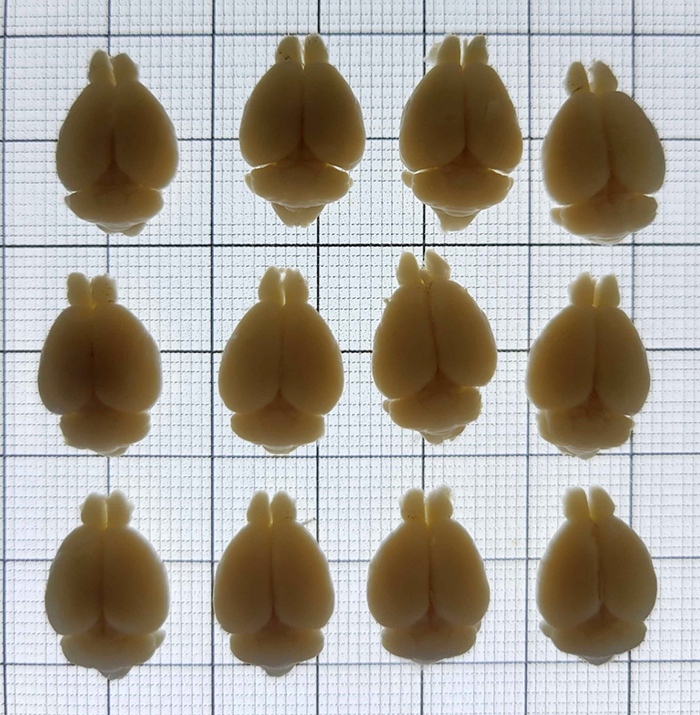
LifeCanvas Brain Clearing Services:
Search available services:
Name
Description
Price
Active Clearing *only*
An accelerated tissue clearing process using the Life Canvas SmartBatch+ system, which enhances the diffusion and uniformity of clearing reagents.
Users must provide their own reagents. This is an active protocol; passive clearing does not require the SmartBatch+.